
Urological Contrast Studies: Intravenous Urography and its Clinical Applications
This article provides an in-depth look at urological contrast studies, with a primary focus on intravenous urography (IVU). It explains how IVU, a commonly used diagnostic tool in urology, works by injecting a contrast medium into the patient’s bloodstream, which then passes through the urinary tract and allows for detailed visualization on X-ray images. The article explains how the procedure typically involves capturing a series of X-ray images from the immediate aftermath of the contrast injection up until the bladder is filled with contrast, around 20 minutes later. The clinical significance of IVU, including its ability to highlight potential abnormalities such as structural anomalies, urinary stones, and tumors, is discussed. The piece underscores the enduring importance of IVU in diagnosing, managing, and monitoring a variety of urological conditions.
Urological Contrast Studies
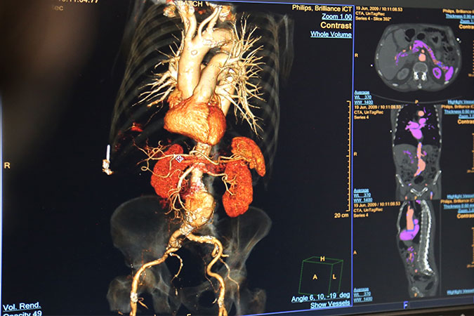
Urological contrast studies are essential diagnostic tools in the field of urology, providing vital insights into the structure and function of the urinary tract. One of the most commonly performed urological contrast studies is intravenous urography (IVU), also known as intravenous pyelography (IVP).
Intravenous urography is a technique that involves the injection of a contrast medium into the patient’s bloodstream. As the kidneys filter this medium from the blood, it passes through the urinary tract, allowing for its detailed visualization on X-ray images.
An IVU procedure typically commences with the intravenous injection of the contrast medium. A series of X-ray images is then captured at different time points, beginning immediately after the contrast injection and extending up to approximately 20 minutes later when the bladder is filled with the contrast medium.
This radiographic series provides a time-lapse visualization of the contrast medium’s passage through the urinary tract, highlighting the kidneys, ureters, and bladder. It allows for the assessment of the anatomy and function of these structures and provides information about the retroperitoneal region and other structures that may impinge upon the urinary tract.
Abnormal findings may include structural anomalies, urinary stones, tumors, or obstructions in the urinary tract. In addition to visualizing the urinary tract, an IVU can also shed light on other aspects of renal function, such as the rate of contrast excretion, which can be informative in patients with suspected kidney disease.
In summary, intravenous urography remains a cornerstone in the evaluation of urinary tract disorders. By providing high-resolution images of the urinary tract and allowing for real-time assessment of renal function, IVU plays a significant role in diagnosing, managing, and monitoring a broad range of urological conditions.