Computed Tomography: Techniques, Interpretation, and Clinical Applications
This article offers a comprehensive overview of Computed Tomography (CT), an advanced medical imaging technology. It discusses the typical presentation and interpretation of CT images, emphasizing the significance of understanding image orientation for accurate diagnosis. The use of oral and intravenous contrast media to enhance the differentiation of anatomical structures and assess vascular patterns is explained, along with the importance of timing in image acquisition for different phases of contrast enhancement. The article further highlights the advantage of CT scanning’s adjustable gray scale, which allows for optimal visualization of various tissues. It recognizes the indispensable role of CT in modern medicine for its capacity to provide high-resolution, cross-sectional images that aid in diagnosing and managing a wide range of medical conditions.
Techniques, Interpretation, and Clinical Applications
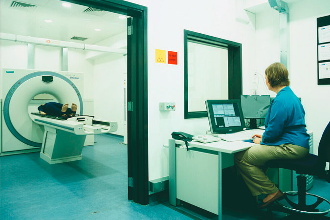
Computed Tomography (CT), also referred to as computerized tomography, represents a significant advancement in medical imaging technology. By harnessing the power of X-ray and computer technologies, CT provides physicians with high-resolution, cross-sectional images of the body’s organs and structures.
The manner in which CT images are presented is crucial for accurate interpretation. Most images are obtained in the axial plane, meaning they’re viewed as if the observer is looking from the patient’s feet towards their head. Therefore, in this standard orientation:
- The right side of the patient corresponds to the left side of the image, and
- The topmost part of the image is anterior (front).
To better differentiate between bowel loops and other abdominal organs and to assess the vascularity of normal anatomical structures, many patients are given both oral and intravenous contrast media. The timing of image acquisition after contrast administration is vital. The earlier the images are obtained post-injection, the more arterial enhancement is seen. As time elapses between contrast injection and image acquisition, venous and equilibrium phases are visualized.
A significant advantage of CT scanning lies in its ability to adjust the gray scale to visualize various tissues effectively. By altering the window settings and window centering, physicians can obtain specific information about different structures, ranging from bones and soft tissues to visceral organs.
With its capacity for detailed, cross-sectional imaging, CT has become an indispensable tool in diagnosing and managing a wide range of medical conditions. It provides physicians with invaluable insights into the structure and function of the body’s internal organs, aiding in accurate diagnosis and informed treatment planning.