Single Photon Emission Computed Tomography: An Overview and Its Clinical Applications
This article provides an overview of Single Photon Emission Computed Tomography (SPECT), a critical medical imaging technique. It outlines the basic principle behind the technique, including the use of gamma rays emitted from injected radionuclides and a rotating gamma camera. The article highlights the broad applicability of SPECT in diagnosing various diseases with specific focus on its utilization in cardiology for diagnosing coronary artery disease and in orthopedics for identifying bone fractures. It concludes by discussing the judicious use of SPECT, considering the exposure to ionizing radiation, and its potential future enhancements.
Single Photon Emission Computed Tomography
Single Photon Emission Computed Tomography (SPECT) represents a significant milestone in the realm of medical imaging. By leveraging the power of advanced gamma-ray detection, this technique has broadened the horizon of disease diagnosis and management.
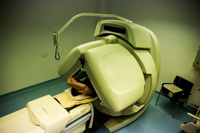
SPECT works through the detection of gamma rays emitted from the decay of injected radionuclides. These radionuclides, commonly being technetium-99m, iodine-123, or iodine-131, are introduced into the patient’s body. As these substances decay, they emit gamma rays, which are then detected by a specialized gamma camera.
This camera rotates 360 degrees around the patient, capturing data from all angles. Advanced computer algorithms then process this data, generating a 3D image of the area under investigation. This results in a comprehensive picture that provides both anatomical and functional information about tissues and organs.
Among the myriad of applications, SPECT has proven particularly valuable in diagnosing a wide range of disease conditions. One of the most significant uses of SPECT is in the field of cardiology, specifically for diagnosing coronary artery disease. The imaging modality allows physicians to visualize the blood flow to the heart muscles, enabling the detection of areas with reduced blood flow indicative of possible coronary artery disease.
In addition to cardiology, SPECT plays a critical role in orthopedics, particularly in diagnosing bone fractures. Traditional X-rays might miss hairline or stress fractures, but SPECT can identify areas of increased bone metabolism indicative of a fracture.
SPECT represents a powerful tool in the physician’s diagnostic arsenal, offering high-resolution, 3D imaging that can aid in the diagnosis and treatment of a variety of health conditions. However, it’s essential to remember that like all imaging modalities, SPECT should be used judiciously and only when necessary, considering the exposure to ionizing radiation. The ongoing evolution of SPECT technology promises to enhance its efficacy, safety, and accessibility, affirming its place in the landscape of medical imaging.