
Shedding Light on the Inside: The Evolution and Impact of Computed Tomography
“Shedding Light on the Inside: The Evolution and Impact of Computed Tomography” is an article that explores the transformative influence of computed tomography (CT) in medical imaging since its invention in the 1970s. The piece provides a comprehensive understanding of how CT scans work, from patient positioning to the capture and processing of images. The article also highlights the numerous advancements in CT technology and its broad applications in diagnosing various health conditions and guiding interventions. Lastly, it underscores the role of CT scans in advancing medical research and improving our understanding of human anatomy and disease pathophysiology.
The Evolution and Impact of Computed Tomography
The invention of computed tomography (CT) in the 1970s by Sir Godfrey Hounsfield marked a significant milestone in the history of medical imaging. For his groundbreaking work, Hounsfield was awarded the Nobel Prize in Medicine in 1979. This monumental achievement set the stage for a technological evolution that has had profound implications for the diagnosis and treatment of a wide range of health conditions.
Computed tomography revolutionized the field of medical imaging by providing clinicians with the ability to visualize the human body in a series of detailed cross-sectional ‘slices’. Before the advent of CT scanning, imaging techniques could only provide a limited, two-dimensional view of the body’s internal structures. In contrast, a CT scan creates a comprehensive three-dimensional image that offers a far more detailed and accurate picture of the body’s interior.
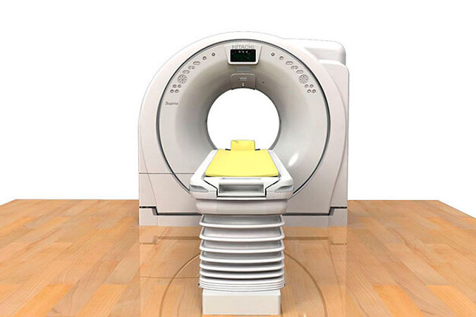
The CT scanning process involves a patient lying on a bed within the CT scanner, a donut-shaped machine. An X-ray tube within the scanner then rotates around the patient’s body, capturing multiple images from various angles. Each of these ‘slices’ offers a detailed view of the patient’s internal structures from a specific angle. However, the true magic of computed tomography lies in the sophisticated algorithms employed to process these images.

The data collected by the CT scanner is processed by a computer, which performs complex mathematical transformations to create a comprehensive, three-dimensional image. This image can then be studied from multiple angles, offering unparalleled insight into the body’s internal structures.
From its inception in the 1970s to the present day, CT technology has seen numerous advancements. Modern CT scanners are faster, more precise, and more versatile than their early counterparts. Today, CT scans are used to diagnose a wide range of conditions, from brain injuries and cancers to cardiovascular diseases and musculoskeletal disorders. They can even guide interventions such as biopsies and drainage procedures.
In addition to its role in diagnosis and treatment, computed tomography has also contributed significantly to medical research. By providing detailed images of the body’s structures, CT scans have deepened our understanding of human anatomy and the pathophysiology of various diseases.
In conclusion, computed tomography represents one of the most significant advancements in the history of medical imaging. This technology has not only enhanced the precision of medical diagnosis and treatment but has also expanded our understanding of the human body. As CT technology continues to evolve, its impact on the field of medicine is set to grow even further.