Magnetic Resonance Imaging: Unraveling the Hidden Depths of the Human Body
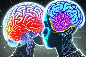
“Magnetic Resonance Imaging: Unraveling the Hidden Depths of the Human Body” delves into the complex and fascinating world of MRI technology. From the physics of proton alignment to the nuanced differences between T1 and T2-weighted images, this article comprehensively explores the principles, applications, and advancements in MRI technology. A critical tool in modern diagnostics, MRI provides highly detailed images of the human body, offering invaluable insights in evaluating vascular diseases and understanding the body’s internal structures. As this technology continues to evolve, it opens up new avenues for improving healthcare outcomes.
Unraveling the Hidden Depths of the Human Body
The realm of medical imaging underwent a transformative revolution with the advent of Magnetic Resonance Imaging (MRI). Initially described in 1946 for determining the structure of complex molecules, the technological and conceptual evolution of MRI has brought about a profound change in how we understand and visualize the human body’s internal structures.
MRI operates based on an intriguing principle: it utilizes the hydrogen nuclei found within water molecules, abundant in nearly all biological tissues, as a diagnostic tool. These protons within the patient’s hydrogen nuclei can be visualized as tiny bar magnets, each with its unique orientation. When a patient is placed within a powerful magnetic field, these ‘bar magnets’ align themselves accordingly.
The real magic happens when a pulse of radio waves is passed through the patient. These waves deflect the aligned ‘magnets’, and as they gradually return to their original state, they emit small radio signals. The strength, frequency, and the return time of these pulses create a distinctive signal. A sophisticated computer system interprets these signals to generate a comprehensive and detailed image of the internal anatomy.
One of the many wonders of MRI technology is the ability to adjust the sequence of pulses, enabling us to assess different properties of the protons, commonly referred to as the “weighting” of the scan. Adjustments in the pulse sequence and scanning parameters facilitate the creation of T1-weighted and T2-weighted images. These two distinct imaging sequences provide varying image contrasts, optimizing and accentuating different tissue characteristics.

In a clinical context, T1-weighted images generally show dark fluid and bright fat. For instance, the cerebrospinal fluid (CSF) within the brain appears dark on these images. On the other hand, T2-weighted images present a bright signal from fluid and an intermediate signal from fat, making the CSF appear white in the brain.

Beyond static imaging, MRI also offers the unique advantage of assessing flow within blood vessels, thereby creating complex angiograms of peripheral and cerebral circulation. This property further enhances its diagnostic utility, particularly in evaluating vascular diseases.
To conclude, MRI stands as a testament to the remarkable strides we have made in medical technology. It provides a non-invasive, highly detailed, and dynamic window into the human body’s hidden depths. As we continue to refine this technology, our ability to diagnose, understand, and manage various health conditions will only grow, improving healthcare outcomes for all.