
Radiological Image Interpretation: The Art of Distinguishing the Normal from the Abnormal
This article delves into the intricacies of radiological image interpretation, a critical component in diagnosing pathological changes in tissues across various clinical specialties. The piece explains the importance of understanding how an image is obtained, the technical considerations involved in the process, and the necessity of recognizing normal anatomy and its variations. It also discusses the process of distinguishing abnormal changes that signify pathological conditions from the normal variations in human anatomy. The article emphasizes the role of radiologists in correlating these variations with the patient’s clinical presentation and history. Finally, it acknowledges the continuous advancements in imaging technologies that promise to enhance accuracy and early diagnosis, thereby improving patient outcomes.
The Art of Distinguishing the Normal from the Abnormal
Radiological image interpretation is a cornerstone of modern medicine. In virtually all clinical specialties, imaging is instrumental in diagnosing pathological changes in tissues, assisting physicians in formulating effective treatment strategies.
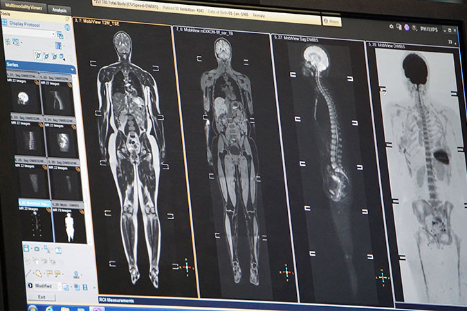
Understanding the process of image interpretation begins with knowing how the image is obtained. Various modalities like X-rays, CT, MRI, ultrasound, PET, or SPECT, all have different techniques and principles of operation. The properties of these techniques dictate the type of information that can be captured, as well as the quality and characteristics of the resulting images.
Central to effective image interpretation is a deep understanding of normal anatomy. Without this knowledge base, distinguishing between normal and abnormal becomes a formidable challenge. Normal anatomy serves as a reference point against which any deviations or anomalies can be identified.
However, it’s also important to recognize that the human body exhibits a significant amount of natural variation. What may appear as an abnormality in one individual might simply be a normal variation in another. This demands an awareness and understanding of the range of ‘normal’ when interpreting images.
Technical considerations also play a critical role in image interpretation. This includes an understanding of how different settings and parameters on imaging machines can affect the resultant images. For example, certain scanner settings may artificially enhance or minimize specific features in an image.
Once equipped with the above knowledge, the process of diagnosing the ‘abnormal’ can begin. Pathological conditions often manifest as changes in tissue structure or function. These changes can present as variations in shape, size, density, or metabolic activity, among others. The task of the radiologist is to identify these variations and correlate them with the patient’s clinical presentation and history.
Radiological image interpretation is both an art and a science, requiring a keen eye, vast knowledge, and an understanding of the principles and technologies used in imaging. The ongoing advancements in imaging technologies promise to continually refine this process, enabling more accurate and earlier diagnoses, ultimately improving patient outcomes.