Gastrointestinal Contrast Examinations: Procedure and Clinical Utility
This article delves into the principles and clinical application of gastrointestinal contrast examinations, diagnostic procedures that utilize high-density contrast mediums for visualizing the gastrointestinal tract. The piece outlines the process of double-contrast studies, explaining how the bowel is insufflated with air or carbon dioxide for enhanced imaging. It touches on the increasing use of endoscopy for upper gastrointestinal imaging but acknowledges the continued importance of double-contrast barium enema for imaging the large bowel. Detailed steps of the double-contrast barium enema procedure, from bowel preparation to image acquisition, are described. The article underscores the vital role of gastrointestinal contrast examinations in diagnosing and managing gastrointestinal conditions, attributing their continued relevance to the detailed anatomical and functional insights they provide.

Procedure and Clinical Utility
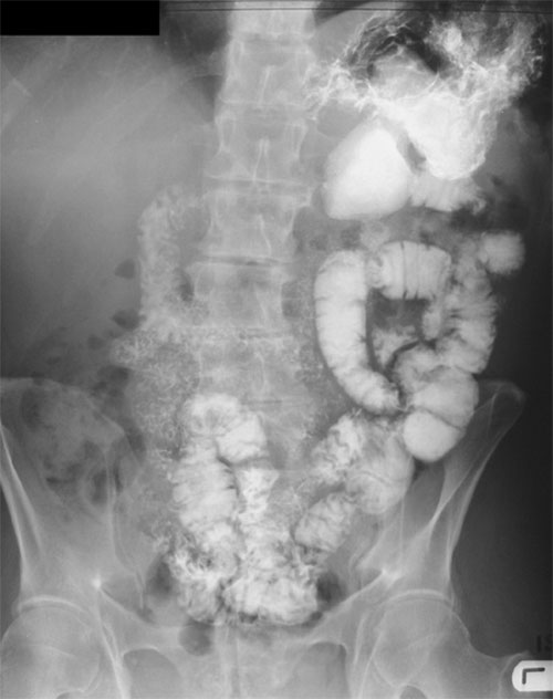
Gastrointestinal contrast examinations are diagnostic procedures used to visualize and evaluate the structure and function of the esophagus, stomach, and intestines. These examinations involve the use of a high-density contrast medium, which is ingested by the patient, to opacify the gastrointestinal tract.
The bowel is often insufflated with air or carbon dioxide to provide a double-contrast study, which can yield detailed images of the gastrointestinal tract. This double contrast can enhance the visibility of the gastrointestinal wall, allowing for the identification of abnormalities such as tumors, ulcers, or inflammation that may not be visible with a single contrast study.
In many countries, upper gastrointestinal imaging has been largely superseded by endoscopy, a procedure that uses a flexible tube with a light and camera to visualize the upper gastrointestinal tract directly. However, imaging of the large bowel, also known as the colon, often still relies on a procedure known as a double-contrast barium enema.

The double-contrast barium enema procedure requires some preparation, which involves using strong laxatives to empty the bowel. During the procedure, a small tube is inserted into the rectum, and a barium suspension is introduced into the large bowel. The patient is then moved into various positions to ensure the barium flows through the entire large bowel.
Once the barium is drained, air is introduced through the same tube to insufflate the large bowel, leaving behind a thin layer of barium that coats the inner lining or mucosa of the colon. This coating allows for the detailed visualization of the mucosa, enhancing the detection of any abnormalities, such as polyps, diverticula, or tumors.
Gastrointestinal contrast examinations, despite the development of other imaging techniques, continue to play a critical role in the diagnosis and management of gastrointestinal conditions. These procedures offer detailed insights into the anatomy and functionality of the gastrointestinal tract, guiding clinicians in delivering effective patient care.


