
Abdominal Radiography: Key Aspects of Procedure and Interpretation
This article explores the concept and clinical utility of abdominal radiography, also known as abdominal X-rays. It describes the typical procedure for obtaining an abdominal radiograph, including the standard anteroposterior (AP) supine position and, when necessary, the erect position for suspected small bowel obstruction. The piece discusses the importance of patient positioning in image acquisition and the key aspects involved in interpreting these radiographs. Emphasis is given to the systematic examination of bowel gas pattern, the detection of free intraperitoneal air, and the identification of any abnormal masses or calcifications. The article concludes by recognizing the critical role of abdominal radiography in diagnosing gastrointestinal disorders, stressing the importance of meticulous interpretation for accurate diagnosis and effective patient management.
Key Aspects of Procedure and Interpretation
Abdominal radiographs, commonly known as abdominal X-rays, provide a non-invasive means of assessing the organs and structures within the abdominal cavity. They can reveal important information about a variety of gastrointestinal disorders, from obstructions and perforations to certain types of inflammation.
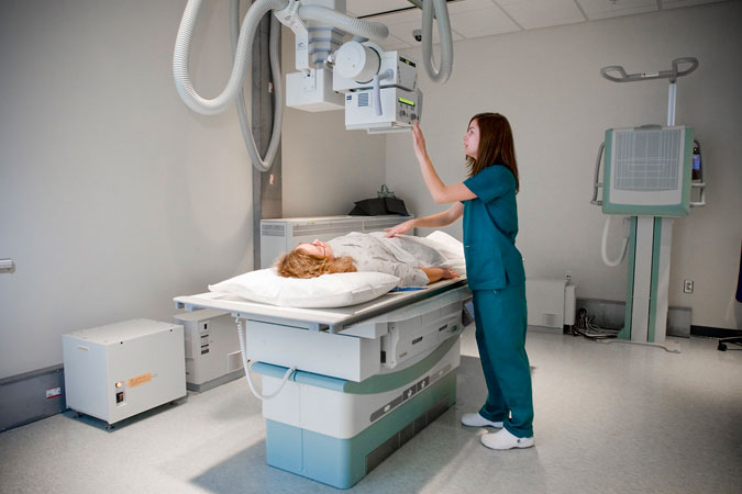
Plain abdominal radiographs are typically acquired with the patient in an anteroposterior (AP) supine position. In this position, the patient lies flat on their back, with the X-ray beam traveling from the front (anterior) of their body to the back (posterior), hitting the X-ray film or detector.
The AP supine position offers several advantages for abdominal imaging. It provides a comprehensive view of the entire abdomen, allows for a more comfortable position for patients who are in pain, and minimizes the radiation dose to sensitive organs like the reproductive organs and bone marrow.
In some cases, an erect abdominal radiograph may be obtained, specifically when there’s a suspicion of a small bowel obstruction. This upright positioning can help visualize free air under the diaphragm, a classic sign of gastrointestinal perforation, and can also help to highlight the levels of fluid and air in the obstructed bowel loops, providing crucial diagnostic information.
Interpretation of abdominal radiographs requires a systematic approach and a solid understanding of abdominal anatomy. It involves examining the bowel gas pattern, looking for any signs of free intraperitoneal air, and identifying any abnormal masses or calcifications.
In conclusion, abdominal radiography is an invaluable diagnostic tool in evaluating abdominal pain and suspected gastrointestinal disorders. The technique and patient positioning in image acquisition play a pivotal role in the diagnostic yield of this imaging modality. As with all radiographs, careful interpretation is key to accurate diagnosis and effective patient management.