Unveiling the Dynamics of Blood Flow: Subtraction Angiography in Focus
“Unveiling the Dynamics of Blood Flow: Subtraction Angiography in Focus” provides an insightful look into the method and significance of subtraction angiography. This piece explains how the technique uses a contrast medium and a sequence of images, combined with digital processing, to visualize blood flow, remove obstructions like bones and tissues, and uncover vascular diseases. Highlighting the transformative impact of this technology in diagnosing and treating vascular conditions, it emphasizes how this procedure has been streamlined thanks to advancements in digital imaging.
Unveiling the Dynamics of Blood Flow: Subtraction Angiography in Focus
Subtraction angiography represents a significant advancement in medical imaging technology. Fundamentally, this procedure involves the introduction of a contrast medium into a patient’s vascular system, followed by a sequence of image captures tracking the contrast’s journey through the arteries and veins.
The magic of subtraction angiography lies in its ingenious image processing method. Initial images are taken before the contrast injection and are digitally inverted to create negatives. Once the contrast agent is injected, new images are captured, detailing the flow of the contrast material through the blood vessels.
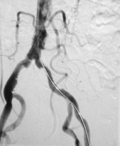
In the subsequent stage, these post-contrast images are added to the negative pre-contrast images. This process effectively cancels out or ‘subtracts’ structures that are consistent in both sets of images – predominantly bones and soft tissues. Consequently, what remains is a clear and detailed view of the contrast agent-filled vessels, free from obscuring bony structures.
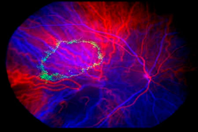
Subtraction angiography offers physicians an enhanced perspective of the cardiovascular system. This powerful visualization tool can reveal the extent and location of vascular diseases such as atherosclerosis and aneurysms. Prior to digital imaging, executing this procedure was arduous. However, with the integration of computers and digital technologies, the process has become straightforward and instantaneous.
Furthermore, the images generated through subtraction angiography can assist in planning surgical or interventional procedures, as physicians can scrutinize the vascular anatomy and pathology in unprecedented detail. This imaging technique has truly revolutionized the way clinicians diagnose and treat vascular conditions, and will continue to evolve with future advancements in imaging technology.