Plain Radiography: The Basic Building Block of Diagnostic Imaging
This article delves into the principles and workings of plain radiography or X-ray imaging, explaining how X-rays interact with different tissues in the body and the subsequent impact on photographic film exposure. It also discusses advancements to this traditional technique, such as continuous X-ray streaming, which enables real-time visualization of moving anatomical structures. Essential for various diagnostic procedures, including barium studies, angiography, and fluoroscopy, these adaptations of plain radiography highlight its continued relevance in the ever-evolving field of medical diagnostics.
Plain Radiography: The Basic Building Block of Diagnostic Imaging
Plain radiography, commonly known as X-ray imaging, forms the cornerstone of diagnostic imaging in medicine. Despite the advent of more advanced imaging modalities, X-ray imaging retains its crucial role due to its simplicity, speed, and efficacy in diagnosing a variety of conditions. This article will explore the fundamental principles of plain radiography, how it works, and some of its modifications for real-time visualization of moving anatomical structures.
The Science Behind Plain Radiography
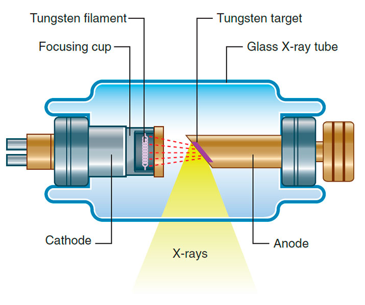
X-rays are photons, which are a form of electromagnetic radiation. They are produced within a complex apparatus called an X-ray tube, which is essentially a type of cathode ray tube. The generated X-rays are then collimated, that is, channeled through lead-lined shutters to prevent them from scattering. The collimated X-rays are directed to the area of the body under investigation.
As the X-rays traverse through the body, they interact with different types of tissues, leading to varying degrees of attenuation, which is the reduction in the energy of the X-rays. This interaction is governed by the principle that different tissues attenuate X-rays differently:
- Air attenuates X-rays only minimally.
- Fat attenuates X-rays more than air but less than water.
- Bone attenuates X-rays the most.
These differences in attenuation result in varying levels of exposure on the photographic film. When the film is developed, areas corresponding to bone appear white because they were exposed to the least number of X-rays due to the high attenuation property of bone. On the other hand, areas corresponding to air appear dark on the film, signifying that these regions were exposed to the highest number of X-rays.
Real-Time Visualization and Beyond
Over the years, various modifications have been made to traditional X-ray techniques to cater to a wider range of diagnostic needs. One of these includes a technique that produces a continuous stream of X-rays from the X-ray tube and collects them on an input screen. This modification allows real-time visualization of moving anatomical structures, making it invaluable in procedures like barium studies, angiography, and fluoroscopy.
Barium studies use barium sulfate, a radiocontrast agent, to visualize the gastrointestinal tract. Angiography uses contrast agents to visualize blood vessels, while fluoroscopy provides real-time moving images of the interior of an organ or a body part, aiding in both diagnosis and treatment.
Conclusion
Plain radiography, a well-established and fundamental diagnostic tool, provides significant insights into the human body. Its versatility and adaptability have allowed it to evolve with the advent of newer technologies, ensuring it remains an essential component of the diagnostic toolkit in modern medicine.