Myotomes: Understanding Muscle Innervation and Functional Testing
Myotomes play a crucial role in understanding the innervation and function of skeletal muscles in the body. They represent specific muscle groups that are innervated by motor fibers originating from a single spinal cord level or a single spinal nerve. Testing myotomes can aid in localizing lesions, identifying nerve involvement, and assessing muscle function. This article delves into the concept of myotomes, their distribution, and their clinical significance in neurological assessments.
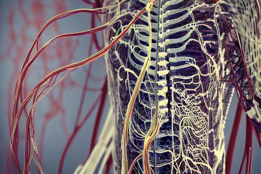
Muscle Innervation
Development and Organization
During embryonic development, somatic motor nerves associated with a specific somite emerge from the anterior region of the spinal cord. These motor nerves, along with sensory nerves from the same level, combine to form a single spinal nerve. Consequently, each spinal nerve carries somatic motor fibers to muscles that originated from the related somite. This arrangement establishes the myotomal pattern, wherein specific muscles are innervated by nerves derived from specific spinal cord levels.
Myotome Distribution
Unlike dermatomes, myotomes are more complex to evaluate as each skeletal muscle in the body often develops from more than one somite. Consequently, these muscles receive innervation from multiple spinal cord levels. However, certain muscle groups are predominantly innervated by specific spinal cord levels, allowing for clinical evaluation and identification of nerve involvement. Understanding the distribution of myotomes helps localize lesions and determine the level of spinal cord or nerve root impairment.

Clinical Significance
Myotomes have significant clinical relevance in diagnosing and assessing motor function. Testing movements at successive joints associated with specific myotomes aids in localizing lesions to specific nerves or identifying the level of spinal cord involvement. By evaluating muscle strength and observing deficits in specific myotomal patterns, clinicians can diagnose and monitor conditions such as peripheral nerve injuries, radiculopathies, and spinal cord lesions. Assessing myotomes is essential in evaluating motor function, designing appropriate treatment plans, and tracking patient progress.
Practical Applications
Understanding myotomes assists healthcare professionals in evaluating muscle strength, identifying specific motor deficits, and determining the extent of motor nerve involvement. Clinical assessments often involve examining muscle groups associated with specific myotomes and evaluating their strength and range of motion. By testing movements at successive joints, clinicians can localize the source of motor impairment, guide targeted therapies, and monitor recovery progress. Myotomal testing is frequently employed in neurological examinations, rehabilitation settings, and sports medicine to assess muscle function and guide treatment interventions.
Myotomes are crucial in comprehending the innervation and function of skeletal muscles in the body. They provide valuable insights into motor nerve distribution, allowing for the localization of lesions and the assessment of motor deficits. By evaluating muscle strength and observing deficits within specific myotomal patterns, clinicians can diagnose and monitor various neurological conditions. Myotome testing plays a vital role in motor assessments, guiding treatment interventions, and tracking patient progress. A comprehensive understanding of myotomes enhances our ability to evaluate and manage motor function, contributing to optimal patient care and rehabilitation outcomes.