Important anatomical terms
This introduction sets the stage for an article that will explore essential anatomical terms used universally in the medical field for clear communication. The article will delve into three key concepts: the “Anatomical Position,” “Anatomical Planes,” and “Terms to Describe Location.” These terms provide a consistent framework for understanding and discussing the structure of the human body, crucial for medical students, healthcare professionals, or anyone interested in human anatomy.
A Fundamental Reference in Human Anatomy
Understanding the complex structure and functions of the human body requires not just knowledge, but also a common language that accurately and universally describes it. To ensure clear communication among healthcare professionals worldwide, certain anatomical terms have been standardized, forming the backbone of any discussion pertaining to human anatomy. These essential terms not only allow precise descriptions of body structures but also facilitate their relative positioning and orientation in a meaningful and universally understood way.
In this article, we will delve into three fundamental aspects of this anatomical language: the “Anatomical Position,” “Anatomical Planes,” and “Terms to Describe Location.” These terms are the basis for all anatomical communication, providing a consistent framework for understanding and describing the intricate layout of the human body. Whether you are a medical student, healthcare professional, or simply an anatomy enthusiast, comprehending these terms is key to navigating and understanding the complex world of human anatomy.
The Anatomical Position: A Fundamental Reference in Human Anatomy
In the world of human anatomy, precision is key. To accurately describe and understand the human body’s structure and function, a common reference point is necessary. This benchmark is known as the ‘anatomical position,’ a standardized stance that forms the basis for all anatomical descriptions and discussions.
Defining the Anatomical Position
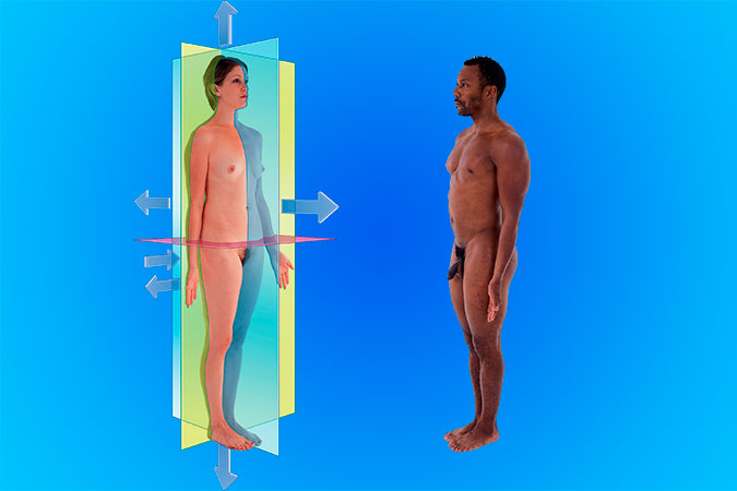
The anatomical position is a universally accepted reference posture used to standardize the description of the human body (Fig. 1). In this position, the body is imagined to be standing upright, with a few distinctive features:
- The person is standing erect, with feet positioned close together. The toes point forward, aligned with the body’s direction.
- The arms are straight and relaxed, hanging down on both sides of the body.
- The palms of the hands are facing forward. In anatomical terms, this is referred to as ‘supination.’ It is essential to note that the fingers are straight, close together, and the thumb pad is turned 90° to the pads of the other fingers, distinctively separate from the rest.
- The head is held high, with the face directed forward. The mouth is closed, and the facial expression remains neutral. Notably, the rim of the bone under the eyes, also known as the infraorbital rim, is in the same horizontal plane as the top of the ear’s opening, referred to as the external auditory meatus.
- The eyes, open and alert, are focused on something in the distance. This forward gaze aligns the visual axis with the body’s direction.
Importance of the Anatomical Position
Adhering to the anatomical position as a point of reference ensures consistency and precision in the description and understanding of the human body. It provides a universally understood framework that eliminates ambiguity and confusion, especially crucial in medical discussions where precision is paramount.
For instance, when healthcare professionals describe the location of injuries, pathological conditions, or medical procedures, they do it concerning the anatomical position. This precise terminology ensures that the information conveyed is clear, accurate, and consistent, regardless of who is communicating or their geographic location.
In summary, the anatomical position is an essential cornerstone of human anatomy, providing a standardized posture that facilitates accurate description and communication. Its universal acceptance allows healthcare professionals worldwide to discuss and understand the human body’s complexities in a unified and coherent manner. This unified understanding ultimately contributes to better healthcare outcomes, aiding in diagnoses, medical procedures, and treatment plans.
Decoding the Human Body: An Introduction to Anatomical Planes
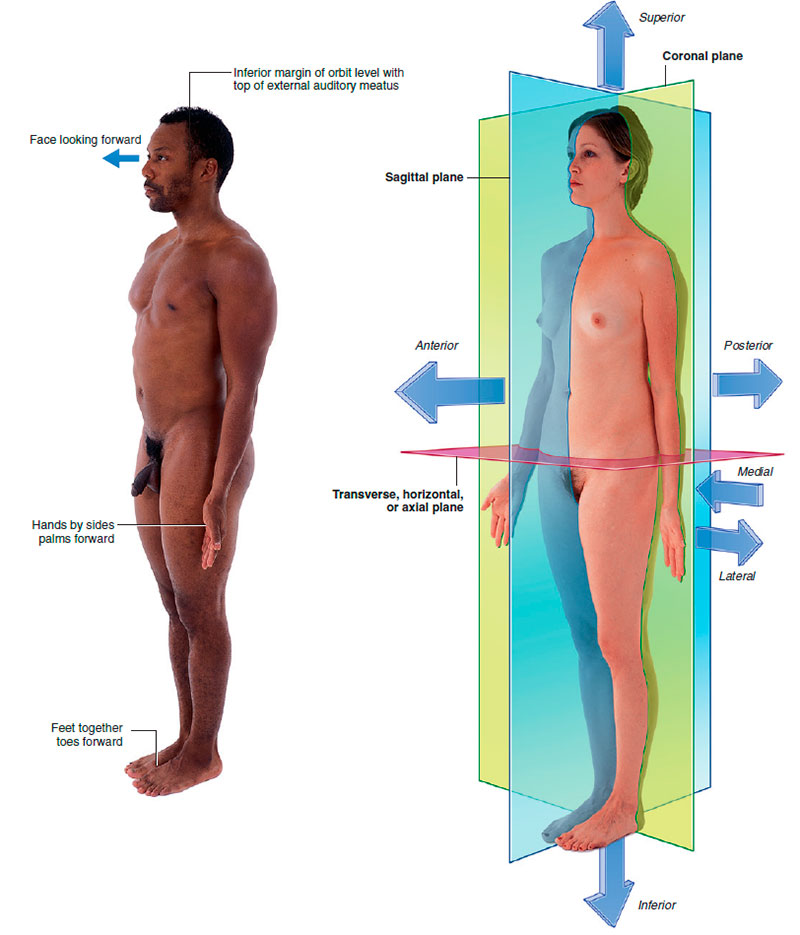
The human body is a complex structure, and understanding its intricacies requires a systematic and organized approach. This is where anatomical planes come into play. Anatomical planes are imaginary flat surfaces that cut through and segment the body, enabling us to visualize, describe, and study its structure and function systematically. There are three principal anatomical planes: the coronal plane, the sagittal plane, and the transverse plane.
The Coronal Plane: Dividing Front from Back
The coronal plane, also known as the frontal plane, runs vertically from one side of the body to the other. It divides the body into anterior (front) and posterior (back) parts (Fig. 1). Named after the Latin word ‘corona,’ meaning crown, the coronal plane runs parallel to the face, as if a crown were placed on the head. When you view the body from a front-on perspective, you are looking along the coronal plane.
The Sagittal Plane: Separating Left from Right
The sagittal plane, similar to the coronal plane, runs vertically. However, it is at right angles to the coronal plane and divides the body into right and left parts. The term ‘sagittal’ is derived from the Latin word ‘sagitta,’ meaning arrow, symbolizing the straight-up-and-down nature of this plane.
There’s a special type of sagittal plane known as the median sagittal plane or mid-sagittal plane. This plane passes through the body’s center and divides it into equal right and left halves. This particular view of the body is commonly used in medical imaging, such as MRIs, as it provides a comprehensive view of many key anatomical structures.
The Transverse Plane: Slicing Top from Bottom
Lastly, we have the transverse plane, also known as the horizontal or axial plane. This plane runs perpendicular to both the coronal and sagittal planes and divides the body into superior (upper) and inferior (lower) parts. The transverse plane is like slicing the body into top and bottom halves as if cutting through a loaf of bread.
Anatomical Planes: Critical to Medical Understanding and Communication
Understanding these three primary anatomical planes is vital to interpreting the structure and function of the human body accurately. They offer a standardized orientation for anatomical descriptions, ensuring clear, consistent communication among healthcare professionals globally.
These planes also have practical applications, particularly in medical imaging techniques like CT scans, MRIs, and ultrasounds. By providing different ‘slices’ or views of the body, these imaging technologies help healthcare professionals diagnose diseases, plan surgeries, and understand more about the body’s inner workings.
In conclusion, the coronal, sagittal, and transverse planes provide a foundational framework for exploring the human body’s complexity. Their standardized descriptions facilitate precise communication and understanding, underscoring their importance in the study and practice of medicine.
Understanding the Human Body: An Introduction to Anatomical Terms for Location
Accurate description and communication about the human body’s structure and function are pivotal in the field of healthcare and medicine. This accuracy relies on a universally accepted set of anatomical terms that describe locations within the body. These terms facilitate precise, clear communication, preventing misunderstandings that could potentially lead to incorrect diagnoses or treatment plans.
In this article, we delve into some of the key anatomical terms that are used to describe location within the human body. We will explore pairs of directional terms, each denoting a relative position within the body. These include: “Anterior (ventral) and posterior (dorsal)”, which describe front and back; “medial and lateral”, which denote positions closer to and farther from the body’s midline; “superior and inferior”, signifying up and down directions; “proximal and distal”, used mainly in the context of limbs; “cranial and caudal”, referring to directions towards the head and tail in the anatomical position; “rostral”, a term specifically used for the orientation in the head; and “superficial and deep”, which describe structures closer to and farther from the body surface.
Whether you are a healthcare professional, medical student, or a curious reader wanting to deepen your understanding of human anatomy, this exploration of anatomical location terms will equip you with a vital tool in the language of medicine. Get ready to navigate the fascinating landscape of the human body with precision and clarity!
Navigating the Human Body: Key Terms to Describe Anatomical Locations
One of the essential facets of studying and understanding human anatomy involves the precise description of the location of various structures within the body. Several pairs of terms are commonly used to denote these relative locations, forming the foundation of communication in the medical and anatomical field. Three such significant pairs of terms are “Anterior (ventral) and Posterior (dorsal)”, “Medial and Lateral”, and “Superior and Inferior”.
Anterior (Ventral) and Posterior (Dorsal)
The terms anterior (or ventral) and posterior (or dorsal) are used to denote the “front” and “back” of the body, respectively. Derived from Latin origins, ‘anterior’ and ‘ventral’ both refer to ‘front,’ while ‘posterior’ and ‘dorsal’ denote ‘back.’
For example, in anatomical terms, the nose is an anterior or ventral structure as it’s positioned towards the front of the body. In contrast, the vertebral column (or the spine) is posterior or dorsal, as it’s located at the back. Thus, the nose is said to be anterior to the ears as it lies in front of them, and the vertebral column is posterior to the sternum (the breastbone), as it lies behind it.
Medial and Lateral
The terms medial and lateral are used to describe the relative position of structures concerning the body’s midline or median sagittal plane and the body’s sides.
‘Medial’ refers to a structure closer to the midline of the body, while ‘lateral’ refers to a structure further from the midline, closer to the sides of the body. For instance, your thumb is lateral to your little finger as it’s farther from your body’s midline. The nose, which aligns with the body’s midline, is medial to the eyes, and the eyes are medial to the ears.
Superior and Inferior
The terms superior and inferior are used to denote the location of structures concerning the body’s vertical axis. ‘Superior’ refers to a structure closer to the top of the head, while ‘inferior’ refers to a structure closer to the feet.
For instance, the head is superior to the shoulders as it’s positioned above them. Similarly, the knee joint is inferior to the hip joint as it’s situated below it.
Conclusion
Understanding these foundational terms – anterior (ventral) and posterior (dorsal), medial and lateral, and superior and inferior – is crucial for precisely describing and understanding the human body’s complex structure. By referring to this framework of terms, healthcare professionals can communicate effectively, ensuring consistent interpretation of bodily locations across the field, leading to more accurate diagnoses, procedures, and treatments.
Anatomical Terms for Precise Localization: Proximal, Distal, Cranial, Caudal, and Rostral
In the world of medicine, anatomy forms the foundation of all clinical practices. The ability to describe precisely where structures are located within the body is of utmost importance. Building on the terms anterior, posterior, medial, lateral, superior, and inferior, we introduce other vital terms used to describe location in the body: proximal and distal, cranial and caudal, and rostral.
Proximal and Distal
When describing the location of structures within the limbs or relative positions of branches along linear structures such as airways, vessels, and nerves, the terms proximal and distal come into play.
‘Proximal’ denotes a location closer to the structure’s origin or point of attachment to the body. On the other hand, ‘distal’ refers to a location farther away from the structure’s origin or point of attachment.
Consider the example of the arm: The hand is distal to the elbow joint as it is farther away from the point where the arm attaches to the body. Conversely, the glenohumeral joint (shoulder joint) is proximal to the elbow joint because it is closer to the arm’s attachment point.
In the context of linear structures like blood vessels, proximal branches occur closer to the system’s origin, while distal branches are located farther away, towards the ends of the system.
Cranial and Caudal
The terms cranial and caudal provide another way to refer to the directions in the body, especially when considering the body in its embryological position.
‘Cranial’ (meaning towards the head) and ‘caudal’ (meaning towards the tail) are often used interchangeably with superior and inferior, respectively. These terms originate from the positions of these parts in four-legged animals, where the cranial or head end is superior, and the caudal or tail end is inferior.
Rostral
The term ‘rostral’ finds its origin in Latin, where ‘rostrum’ refers to ‘beak’. In the context of human anatomy, rostral is used specifically in the head to describe the position of a structure concerning the nose, similar to how an animal’s structure might be related to its beak.
For instance, when discussing parts of the brain, the forebrain is rostral to the hindbrain, meaning it is located closer to the front (nose side) of the head.
Conclusion
Understanding the anatomical terms proximal, distal, cranial, caudal, and rostral, along with the other directional terms, enables clear and unambiguous communication in the medical field. These terms form a common language that healthcare professionals use to deliver effective and precise care, from diagnosis to treatment. As such, knowledge of these terms is fundamental to any person involved in healthcare, from students to seasoned professionals.
The Layers of Human Anatomy: Understanding Superficial and Deep Structures
When navigating through the layers of human anatomy, it becomes essential to distinguish between structures that are closer to the body surface and those nestled more deeply within. Enter the terms ‘superficial’ and ‘deep’ – these provide the foundation for describing the relative location of structures concerning the body’s surface.
Superficial and Deep: Relative Positioning
‘Superficial’ and ‘deep’ are anatomical terms used to describe the relative position of a structure concerning the body’s surface. The term ‘superficial’ refers to a structure situated nearer to the body’s surface. In contrast, ‘deep’ denotes a structure that is located further away from the surface, closer to the core of the body.
Take, for instance, the sternum (the flat bone in the center of the chest) and the heart. The sternum is superficial to the heart, as it is closer to the body’s surface. Similarly, the stomach is deep to the abdominal wall as it is located further inward from the body surface.
Superficial and Deep: Absolute Classification
Beyond their usage in relative positioning, ‘superficial’ and ‘deep’ can also be used in a more absolute manner to define two major regions of the body.
The superficial region of the body refers to everything external to the outer layer of deep fascia, a dense layer of connective tissue surrounding muscles and other internal organs. Structures in this superficial region include the skin, superficial fascia (a looser connective tissue layer beneath the skin), and mammary glands.
Conversely, deep structures are those enclosed by the outer layer of deep fascia. These typically include skeletal muscles and viscera, the large internal organs within the body cavities.
The distinction between superficial and deep also provides a useful framework when assessing injuries. Superficial wounds affect the areas external to the outer layer of deep fascia, while deep wounds penetrate this layer, potentially impacting the enclosed structures.
Conclusion
Understanding and effectively utilizing the anatomical terms ‘superficial’ and ‘deep’ can ensure precise communication in the medical field. These terms provide a universal language that facilitates a clear understanding of the body’s structure, aiding in everything from routine physical examinations to complex surgical procedures. A firm grasp of these concepts forms the cornerstone of comprehensive anatomical understanding, facilitating effective patient care.