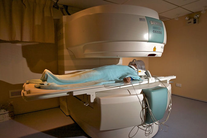
Decoding Disease: Understanding Diffusion-Weighted Imaging
“Decoding Disease: Understanding Diffusion-Weighted Imaging” is an in-depth exploration of the innovative medical imaging technique, Diffusion-Weighted Imaging (DWI). This article delves into the science behind DWI, explaining how it utilizes the principles of Brownian motion to track the movement of water molecules in body tissues, thereby creating detailed images that can differentiate between normal and abnormal tissues. DWI’s invaluable role in detecting diseases, such as tumors and infarcts, and its promising potential in monitoring therapeutic responses are discussed, underscoring its significance in modern medical diagnostics.

Understanding Diffusion-Weighted Imaging
In the realm of medical imaging, advancements in technology have allowed physicians to visualize the human body’s internal structures with increasing precision. One such revolutionary technique is Diffusion-Weighted Imaging (DWI), a type of Magnetic Resonance Imaging (MRI) that provides a distinct lens into the microscopic movement of water molecules within tissues. This insightful methodology enhances our ability to delineate normal tissue from pathological areas, proving particularly beneficial in diagnosing conditions such as tumors and infarcts.
Understanding the Science behind DWI
The principle behind DWI is rooted in the phenomenon of Brownian motion, named after the scientist Robert Brown who first observed the erratic, random movement of particles suspended in fluid. In the context of DWI, Brownian motion refers to the natural movement of water molecules within body tissues.
Under normal conditions, water molecules freely diffuse in the extracellular spaces, while diffusion is more restricted within cells. This dichotomy in motion creates a unique signature of water movement for different tissue types, which can be exploited to create detailed images.
DWI uses this variability in diffusion patterns to generate contrast in MR images. By applying a series of gradient magnetic fields, DWI accentuates the impact of Brownian motion on the signal. Consequently, areas where water molecules move freely (high diffusion) appear dark, while areas where water movement is restricted (low diffusion) appear bright.
DWI in Detecting Disease
In disease states such as tumors and infarcts, the balance of water movement is disrupted. Tumors, for instance, exhibit a higher cell density compared to normal tissue, leading to an increase in intracellular fluid. Consequently, there is a higher proportion of water molecules confined within cellular boundaries, which restricts their movement. Similarly, in the case of infarcts, the death of tissue and cells causes an influx of water into the intracellular compartment.
These scenarios result in overall increased restricted diffusion, making the affected areas appear brighter on a DWI scan. This ability to detect subtle changes in tissue composition makes DWI a powerful tool in identifying abnormal tissues and enhancing the accuracy of diagnoses.
Real-World Applications of DWI
In clinical practice, DWI has become invaluable, particularly in the realm of neuroimaging. DWI can detect acute cerebral infarction (stroke) within minutes of symptom onset, much earlier than conventional imaging techniques, thereby enabling swift intervention. Similarly, DWI is employed to differentiate benign from malignant lesions, thanks to its sensitivity to the cellularity of tumors.
Beyond its prowess in identifying diseases, DWI also offers potential in monitoring therapeutic responses. For example, following successful treatment, the cellularity of a tumor may decrease, reflected by changes in diffusion patterns on DWI.
In Conclusion
Diffusion-Weighted Imaging, with its ability to unveil the microstructural changes in tissues, represents a breakthrough in medical imaging. By harnessing the physics of water molecule movement, DWI opens a window into the inner workings of the human body at the cellular level. As we continue to explore this technique’s capabilities, we can expect DWI to play an increasingly prominent role in disease diagnosis, management, and monitoring.


