An Extensive Examination of Synovial Joints: The Cornerstones of Human Movement
This article provides a comprehensive review of synovial joints, emphasizing their structure, functionality, and significance in human movement. It details the characteristic features of synovial joints, including hyaline cartilage, the joint capsule (with its synovial and fibrous membranes), and additional structures such as articular discs and fat pads. By shedding light on these integral elements and their respective roles in maintaining joint stability and facilitating movement, the article underlines the importance of understanding these structures for the medical community, particularly in the field of orthopedics.

Synovial Joints
Synovial joints, integral components of the human skeletal system, are known for their pivotal role in facilitating movement. Characterized by the presence of an articular cavity, these joints feature a unique architecture and complex functionality, all of which will be thoroughly explored in this article.
The Basics of Synovial Joints
Firstly, it’s important to establish what defines a synovial joint. In essence, a synovial joint is a connection between two or more skeletal elements, separated by a narrow articular cavity. This cavity is filled with synovial fluid, which serves to reduce friction and ensure smooth articulation between the connected elements.
Unique Structural Components of Synovial Joints

Synovial joints are distinguished by several characteristic features:
Hyaline Cartilage
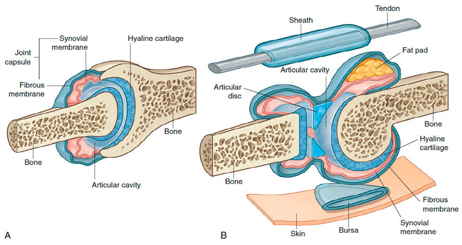
In synovial joints, the articulating surfaces of the skeletal elements are covered by a layer of hyaline cartilage. This cartilage ensures that the bony surfaces do not directly contact each other, reducing the risk of wear and tear. This cartilage layer is also the reason why, when viewed on radiographs, a substantial gap seems to separate the adjacent bones. The cartilage is more transparent to X-rays than bone, resulting in this apparent gap.
Joint Capsule
Synovial joints feature a joint capsule, composed of an inner synovial membrane and an outer fibrous membrane:
- Synovial Membrane: This membrane attaches to the joint surface margins at the point of interface between the cartilage and bone. It is responsible for enclosing the articular cavity. The synovial membrane is highly vascularized, producing synovial fluid, which lubricates the articulating surfaces. In some cases, this membrane also forms synovial bursae or tendon sheaths outside the joints, reducing the friction between various structures, such as tendons and bones.
- Fibrous Membrane: Made of dense connective tissue, this membrane surrounds and stabilizes the joint. In some instances, it may thicken to form ligaments, which provide further joint stability.
Additional Structures
Many synovial joints also feature other structures within the area enclosed by the joint capsule or synovial membrane, such as articular discs, fat pads, and tendons:
- Articular Discs: Usually composed of fibrocartilage, these discs play a crucial role in absorbing compression forces, adjusting to changes in joint surface contours during movement, and increasing the joint’s range of motion.
- Fat Pads: Located between the synovial membrane and the capsule, fat pads move into and out of regions as joint contours change during movement. They serve as additional protective structures.
In Conclusion
Synovial joints, with their unique structural features and complex functionalities, showcase the marvels of human biomechanics. Their role in facilitating a wide range of movements, while maintaining stability, is a testament to their intricate design. A deeper understanding of these components is not only fundamental to the field of orthopedics, but also instrumental in advancing treatment strategies for numerous joint-related conditions, ultimately improving the quality of life for countless individuals.


